About 10 percent of the genes in the human genome encode transcription factors. Transcription factors are proteins that bind to DNA, controlling gene expression. Notably, they control the precisely-timed differentiation of stem cells into specialized tissues during the development of organisms.
In an article published online in the journal Nature this week, Marius Wernig and colleagues at Stanford University demonstrate that the expression of only three transcription factors, that isAscl1, Pou3f2 and Myt1l, is needed to convert cultured fibroblasts, that is undifferentiated connective tissue cells, into nerve cells (Vierbuchen and others, 2010). The cells were shown to produce action potentials, i.e. the electrical discharges that propagate along their output processes called axons, and formed functional connections, known as synapses, with other nerve cells.
Although the conversion has been successfully carried out only in a dish with developing mouse tissue and only the most essential nerve cell functions have been demonstrated, the method does not utilize stem cells and still may bear great potential for therapies, seeking to replace lost nerve cells in neurodegenerative diseases, e.g. Parkinson's disease.
Addendum
If you are considering stem cell therapy, you may find the information on the International Society for Stem Cell Research site helpful (07/26/10).
Last Wednesday, Jan. 12, 2010, an earthquake of magnitude 7.0 shook the island nation of Haiti at 16:53 local time. The epicenter of the quake was located only about six miles from the nation's largest city and capital Port-au-Prince, where an estimated 2.5 million people live. The temblor collapsed buildings within seconds, burying hundreds of thousands under the rubble. The government was not able to mount any first response of note. No rescue teams showed up on the scene. No heavy equipment was rushed in to help free the trapped from under the rubble. People shoveled with bare hands. The police was not to be seen. A human catastrophe of apocalyptic proportions was going to unfold.
In spite of the ensuing international rescue effort and the medical assistance that has begun to reach the country, hundreds of thousands are believed to have died. Many more are feared to succumb to their injuries in coming days. Many destroyed dwellings seem flimsily erected with cinder blocks and mortar. Reinforced concrete structures with multiple floors were not quake-proof.
The tumbling of concrete walls and ceilings caused countless blunt-force traumas and open fractures among the survivors. People with such injuries need immediate medical attention, including surgery. Only few help organizations were prepared for such assistance in the immediate aftermath of the quake. International medical teams including surgeons from Médecins Sans Frontières and Cuba were on location and are to be commended for their decisive action in the first hours after the quake. Meanwhile, aid is pouring in. The relief effort has become more coordinated. Field hospitals are being set up. Food and water are distributed. Security forces can be seen in action.
Hopefully, the basic needs of the survivors will be met in the coming days. It is clear that the horrific consequences of this catastrophe are not entirely natural. Rather, they seem in good part man-made. That is, the Haitian government has failed colossally in its role as a protector of its citizens. Numerous recent accounts of international volunteers who have been looking after the most profound needs of the country's impoverished population bear witness to the enduring insufficiency of medical care and the endemic shortages of the most basic medicines like antibiotics (e.g. Joani in Haiti). Hospitals had been strained at best before the disaster and have now been rendered completely inadequate. The temblor has laid bare once more the societal ills of this aggrieved nation.
I am ill-prepared to discuss the economical, social, and political reasons for Haiti's failure. The Duvaliers and their somewhat more benign successors certainly did not contribute much to lead the country out of its misery. The international community and the independent organizations providing aid to Haiti may wish to re-assess the ways they can possibly help build a more efficient infrastructure and social services in this country in the future.
Watching the horror take its course, I kept wondering whether we cannot find low-cost indicators to predict an earthquake, if only immediately before the temblor strikes. Even if long-term predictions similar to those advising us of the strength and the path of a hurricane do not seem feasible for earthquakes to date, a few more seconds of warning may make a huge difference.
A day after the quake, CBS News posted a video covering a road lined with a row of condominiums on the far side at the time the event.
The 35-second long footage was captured by a rigidly-mounted camera overlooking an intersection. Twenty seconds into the footage, a series of very strong vertical displacements of the ground brings the first building down, raising a huge plume of dust. Could we have noticed early signs in the video warning us that a strong quake was about to happen?
Though the quality of the video is not high fidelity, examining the pictures closely provides a few insights. We need to focus on the roof line of the condos first and then on the utility pole standing on the near corner of the intersection.
A subtle distortion of the roof line announces the first vertical jolt 12 seconds into the footage. Subsequent, arrhythmic displacements gain strength within the next 3 seconds. At 16 seconds, we see a distinct solitary vertical jolt followed by a pause of a second. Then the ground begins to violently shake up and down at precise periodicity. Note that the utility pole sways in elliptical counter-clockwise rotation. At 20 seconds, after six strong vertical jolts in four seconds, the front condo pancakes. Nothing in the first 12 seconds of movie foretells the impending disaster.
I found information on earthquakes pertinent for the interpretation of our observations in the movie on L. Braile's web page at Purdue University's Earth & Atmospheric Sciences. Three types of seismic ground waves that propagate parallel to the earth's surface appear to come to bear in the movie.
Compressional P waves are known to travel fastest and arrive first. They are longitudinal waves, shaking the soil particles back and forth in the direction of propagation like lined-up billiard balls. P waves cause comparably small vertical ground displacement. The next to arrive are shear or S waves. This type of wave propagates transversely with soil particles jolted up and down at larger amplitude than P waves. At last, Rayleigh or R waves arrive. These waves move soil particles in the direction of and vertical to propagation, causing the ground to rotate elliptically as the utility pole in the movie convincingly demonstrates. R waves brought the condo in the movie down. In addition, a fourth type of wave, Love or L waves, has been recognized. I could not detect any manifestation of L waves in the movie.
Braile exemplifies the different wave types in figure 7 on his web page. The figure shows seismograms recorded in Peru during a magnitude-6.5 quake off the Pacific coast in 1998. The trace representing the displacement in the z-direction (vertical) at the bottom of the figure is most applicable to our observations in the movie. The difference in time between the onset of the P and the S waves multiplied by 8 provides a rough estimate of the distance between recording site and the epicenter of the quake in kilometers. In the Peruvian example the seismic recorder was more than 1000 kilometers removed from the quake's epicenter. The S waves lagged the P waves about 7 minutes. By contrast, according to the United Nations Office for the Coordination of Humanitarian Affairs the distance between the epicenter of the quake in Haiti and downtown Port-au-Prince was only 10 kilometers. This leaves just 2 seconds between the arrival of the P waves and the S waves in the movie. If the P waves were strong enough to cause the earliest vertical displacements we noted, the inhabitants had seven seconds to vacate the condo complex before it crumbled. If the P waves were too weak to cause any visible displacement and the first detectable jolts were caused by S waves, we may add another two seconds according to the rule of thumb explained above. We would have had nine seconds to leave the building, after we first felt the ground shake. This may sound short. However, let us not forget that the world's fastest runner, the Jamaican Usain Bolt, covers 109 yards in that time.
Would we have recognized that the first mild vibrations were the harbingers of a temblor of catastrophic force arriving shortly? Animals may possess a better sense of such danger than humans. In another contribution, CBS News broadcast a segment interviewing geologists who foresaw a major quake near Haiti a couple of years ago. They just could not predict when disaster would strike. In that broadcast, a video from a different quake shows a dog in an office that unequivocally recognizes the imminent catastrophe before any human. The dog takes flight about 5 seconds before the room begins to shake violently.
Animals are known to behave conspicuously in advance of an earthquake, and researchers have tried to associate specific types of behavior with the event (Mott, 2003). To date, the findings of these studies have not yielded any consistent predictors. However, I would not be surprised, if burrowing animals, that are particularly attuned to the processing of low frequency vibrations, would sense earthquakes early, displaying distinct behaviors that could be useful as warning signs. I have written about such animals in my post entitled "The Quest for the Infrasound Fovea" dated Oct. 12, 2009. In fact, the mammal that survived in the pumice zone after the catastrophic eruption of Mt. St. Helens in 1980 was a tiny whiskered burrowing rodent (Force & Resilience).
Addenda
According to Madeline Brand's segment with the title "Technology Works to Provide Early Quake Warning" broadcast on National Public Radio's All Things Considered today, sophisticated technology exists that permits us to identify a dangerous earthquake early enough to provide minutes of advance warning. However, when we listen to the podcast, we must keep in mind that the warning time dwindles as we near the quake's center. Even if such equipment had been installed in Haiti, we still would have had only seconds to act in Port-au-Prince (01/29/10).
On Saturday, Feb. 27, 2010, 3:34 local time an earthquake of magnitude 8.8 on the Richter scale originated under the Pacific Ocean at a depth of 22 miles 60 miles offshore Chile near Maule. An account of the event is currently being developed on Wikipedia (2010 Chile earthquake).
The temblor was measured fifth strongest on record, inflicting wide-spread, severe damage along the coast of Chile. Ensuing tsunami waves, which topped ten feet in some locations caused additional devastation. To date, the loss of life stands at about 800. Mostly old, historic structures suffered catastrophic failure. Earthquake-resistant construction methods in modern buildings saved many lives in metropolitan areas. By contrast, in Haiti such building practices constituted a rarity (03/03/10).
According to Terry Wade and Fabian Cambero's post entitled "Chile quake-area still shaking, death toll unclear" on Reuters today, the loss of life owing to last Saturday's earthquake off the Chilean coast and the ensuing tsunami is much smaller than believed earlier. The Chilean government now claims that roughly 300 people died as a result of the catastrophe (03/05/10).
In today's post on Reuters with the title "Chileans' quake knowledge saved thousands of lives", Daniel Trotta provides a few more details released by the U.S. Geological Survey about the Chilean quake three weeks ago. A 41-foot jump of the tectonic Nazca plate toward the South America plate triggered the 8.8-magnitude quake, 500 times more powerful than the magnitude-7.0 quake that struck Port-au-Prince six weeks earlier. The Nazca plate usually moves roughly 3 inches per year. In addition to strict building codes enforced in Chile, the low loss of lives compared with Haiti may be attributable to the atypically late arrival of the most destructive jolts after the first noticeable tremors. R and L waves arrived between 20 and 30 seconds after the P waves. In Port-au-Prince, this difference in time was only about 5 to 8 seconds. The Chilean coast was hit by three tsunami waves, surging 26 to 33 feet in some locations. Most lives may have been lost because of the flooding (03/19/10).
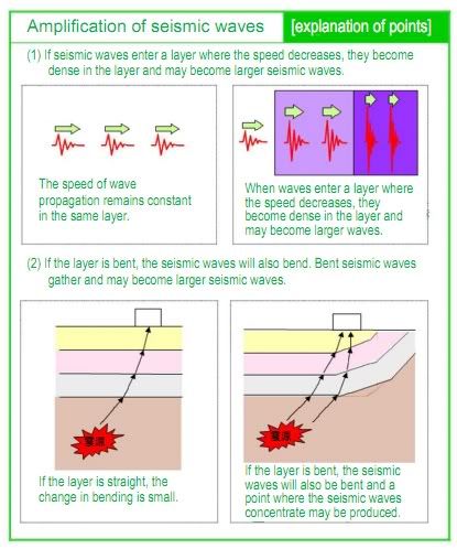
Hough and others (2010) demonstrate in a recently published seismological research study that topographic features around Port-au-Prince, like a steep ridge, must have amplified ground motion by constructive interference during the Haiti earthquake, in addition to the composition of superficial sedimentary layers in low-lying areas. Perhaps the ridge is the best location to detect an impending quake (10/18/10).
Not much progress in a year as this Reuters slide show published today with the title "Haiti's somber anniversary" illustrates (01/11/11).
Nova aired this informative program entitled "Deadliest Earthquakes" on recent advances in understanding the nature of earthquakes on Jan. 11, 2011 (01/30/11).
The philosopher Martin Heidegger reasoned in his treatise "Sein und Zeit" that our sense of time confers our sense of being (Heidegger, 1976). Henry Gustav Molaison could attest to that. In order to relieve Henry from severe bouts of epileptic seizures, a part of his temporal lobes had been removed when he was in his late twenties.
The seizures subsided. However, as an unwanted consequence of the surgery, he had lost essential elements of his memory. He could not remember who he was and had to discover his identity anew every day. One day late in his adult life, Henry was caught curiously examining his face in a mirror. He called out in amazement, "I am not a boy!" In Heidegger's words, Henry Molaison had lost his sense of time and, with that, his sense of being.
As patient H.M., Henry volunteered for scientific research throughout his life, becoming one of the most intensely examined cases in psychology. He willed his brain to research. Yesterday a year ago, he passed away of respiratory complications at the age of 82. On the first anniversary of his passing, the histological processing of his brain was begun at the University of California San Diego. The university's Brain Observatory provides live online footage of the sectioning of the frozen whole brain on a cryotome until Dec. 4, 2009. The brain is cut coronally, that is in the transverse plane. Seventy micrometer-thin sections are collected for histology at equally-spaced intervals. At the speed maintained thus far, we shall reach the site of surgery tomorrow.
The ensuing histological examination of his brain will complete Henry's scientific journey as he willed. The findings may provide important insights into the histochemistry of memory, constituting the keystone in the arch of knowledge that H.M. allowed us to build.
At 5:00 am (CST), we are in the midst of the temporal lobe of H.M.'s brain. If you look closely at the bottom of the brain (ventral, on the left hand side of the observer), you see cauliflower-shaped protrusions on both sides. These are the temporal lobes. There is a void inside the structures. Parts of the hippocampus seem absent (12/04/09).
At about noon, in section 1672 (46,344 micrometers), we can see a cavity in left lingual gyrus (in the hemisphere closest to the observer) (12/04/09).
At 5:00 pm (CST), we are entering the occipital lobes; at 10:00 pm we passed the middle of the occipital lobes (12/04/09).
We should be able to detect signs of nerve cell and nerve fiber degeneration in the brain regions that were connected to the brain tissue that was removed or damaged otherwise during surgery, particularly in the structures of the limbic system. Degenerating nerve fibers can be rendered decades after the damage and ameboid microglia may reside in the affected locations (Miklossy and others, 1991). According to von Monakow's concept of diaschisis, the affected regions can be far-flung and distant from the site of immediate damage, comprising the opposite cerebral and cerebellar hemispheres. That is why H.M.'s whole brain needed to be sectioned, and 2,401 sections were collected (12/05/09).
The footage covering the sectioning does not seem to be available on The Brain Observatory's site at this time. You may find a CBS report broadcast Dec. 3, 2009, instructive (01/01/10):
Brain sections are prepared for histology with two distinctly different methods. One way requires that the tissue is embedded in hot paraffin wax or plastic. The tissue is sectioned at room temperature on a bench top with a microtome or ultramicrotome, respectively. Paraffin embedding is widely used in routine pathology. Plastic is used to embed specimen to be cut into ultra-thin sections for electron microscopy. The other method of tissue preparation requires that the specimen are frozen. Frozen tissue has to be stored and sectioned at temperatures below 0 ℃.
With either method of preparation, the tissue is commonly first soaked in solutions of formaldehyde, glutaraldehyde or a mixture of both. The aldehydes help bond cellular protein, preserving cellular structure during the further processing steps. H.M.'s brain was then soaked in highly concentrated sucrose solutions and embedded in dextran. Sucrose and dextran are sugars. The sucrose prevents ice crystals from forming in the cells when the tissue is frozen. The growing crystals would otherwise burst the cells. The dextran helps preserve the integrity of the sections, preventing tissue from breaking off the perimeter of the specimen during cutting. Once the brain is fixed and cryo-protected, it can be cut into sections that are only tens of micrometers thick on a microtome on which only the stage holding the brain is kept at subzero temperatures. The sections are picked up from the microtome's knife with a brush and can be processed freely floating in solutions containing the substances needed for the chosen histological staining procedure.
There are numerous histological staining procedures. Most comprise chemical reactions. Others involve antibodies that bind specifically to cellular proteins or fluorescent dyes that fill particular cellular components. Among the oldest procedures is the silver impregnation method named after Camillo Golgi, rendering the cell bodies of nerve cells and a good part of their processes (dendrites) and protrusions (dendritic spines). This method helped Ramón Cajal to divide the cerebral cortex into six layers, a distinction that is still used today. Golgi and Cajal were awarded the Nobel Prize for their contributions. The most commonly employed methods, however, are the Nissl stain for cell bodies and the myelin stain that stains the fatty sheeth around nerve cell fibers in the brain's white matter. These methods will certainly be used with some sections of H.M.'s brain. After staining, the sections are mounted on glass slides, dehydrated in solutions of increasing alcohol concentration, embedded in a clear organic solvent-based glue and covered with thin glass coverslips.
The methods of tissue preparation described above cannot be used when the procedures require that fixation-sensitive cellular proteins remain functional or when water-soluble, diffusible substances are to be localized in the tissue. In that case, the tissue must remain unfixed and immersion in sucrose cannot be used. The tissue has to be shock frozen at -70 ℃ and lower, and the sections must be kept frozen during sectioning. To fulfill this need the whole microtome is encased in a refrigerated cabinet. The instrument is then called a cryostat. Ten or 20 micrometer-thick sections are cut and picked up on chilled glass slides and quickly dried on a hotplate outside the cabinet. The subsequent procedures must consequently be carried out on the slide-mounted sections. This method is used for the autoradiography of diffusible radioactively-labeled tracers. For autoradiography, the sections are exposed to x-ray films or radiation-sensitive semiconductor chips that produce gray-scale images of the distribution of the tracer in the sections. The optical densities in the images called autoradiograms are proportional to the concentration of the tracer in the tissue and can be measured.
In order to trace metabolites in whole bodies, Sven Ullberg and colleagues at Uppsala University, Sweden, engineered a microtome sufficiently sturdy to cut thin sections through the whole body of small animals. The microtome was placed it in a chest freezer with windows in the lid (Larsson and Ullberg, 1981). The instrument is called a whole-body cryotome and was commercially available from the Swedish company LKB. I was privileged to use a LKB cryotome 30 years ago for functional imaging studies (Melzer, 1984). The picture below shows a whole rat embedded length-wise in dextran and frozen in liquid nitrogen at -173 ℃. The dextran block with the rat is mounted on the sliding stage of the cryotome for sectioning.
The block of dextran with the rat resembles that with H.M.'s brain in the footage of the Brain Observatory which misled me initially. But as I pointed out, H.M.'s brain was fixed and protected with sucrose. Only the microtome stage had to be cooled. No freezer chest was needed. Jacopo's hands stayed warm.
One great advantage of whole body sectioning is that the sections render the organs in their actual position inside the body. The picture below shows a the block surface of the head of a sandrat embedded in dextran and cut transversely at the level of the midbrain.
The structure at the center is the brain. The glassy cavities on both sides of the brain are the inner and middle ears. The middle ear cavities of sandrats are remarkably enlarged compared with other rodents, serving as a low pass filter. The animal had been exposed to sound for functional imaging. Brain activation images are shown in my post entitled "The Quest for the Infrasound Acoustic Fovea" and dated Oct. 12, 2009.
Since the brain was preserved accurately as it was situated in the scull, we were able to pinpoint brain structures with stereotaxic precision, a goal that may be achieved with structural magnetic resonance imaging today. That method did not exist then.
In addition to functional imaging, fresh tissue tissue sections permit us to examine the distribution of radioactively-labeled ligands that bind to specific receptors on cell surface or probes that identify sequences of genetic code. These procedures can not be applied to the sections of H.M.'s brain. However, even with the histological techniques available a treasure of knowledge will be uncovered from the brain that Harvey Molaison kindly willed.
In the past three decades most departments of anatomy have been transformed into departments of cell biology for good reason. However, gazing through a microscope at the multitude of diversely shaped nerve cells in a Golgi-stained section through cerebral cortex still remains as fascinating as gazing through a telescope at the myriad of stars in the night sky. The neuroanatomical methods that make this experience possible resemble in many ways forms of art, though rather of an artisan than of an artist. Perhaps, the work on Harvey's brain will rekindle interest in this type of expertise, paving the way for a renaissance of neuroanatomy (01/02/10).
Melzer P (1984) Whole head sectioning in [3H] deoxyglucose mapping of auditory responses in gerbils. Brain Res Bull 12:331-334.
Miklossy J, Clarke S, Van der Loos H (1991) The long distance effects of brain lesions: visualization of axonal pathways and their terminations in the human brain by the Nauta method. J Neuropathol Exp Neurol 50:595-614.