advertisement

Pages

Showing posts with label neural networks. Show all posts

Friday, December 30, 2011

Tepco 東京電力 and the Toad 蟾蜍 : Neuroethological Reflections on the Failure of A Human Enterprise


“During the week from March 11, I thought several times that I would die.”Director of the Fukushima Dai-ichi Nuclear Power StationMasao Yoshidain his interview with Asahi Shimbun published online Nov. 13, 2011 under the headline "Nuke plant director: 'I thought several times that I would die.'"
“It was a crucial moment when I wasn't sure whether Japan could continue to function as a state.”Prime Minister of Japan His Excellency Naoto Kan published online in The Japan Times under the headline "Tokyo faced evacuation scenario: Kan" published online Sep. 19, 2011.
Looking back on 2011, the year reminded us ever more strongly of our limited power of comprehending risk. The two statements above epitomize the most consequential technological failure of 2011.

The islands of Japan are home to a variety of toads. The video below shows a large specimen of Bufo japonicus formosus or アズマ - 比企ガエル in Japanese. I suppose the people of Japan are well acquainted with this creature.


Toads are not appreciated enough. Important lessons can be learned from the simple behaviors these creatures afford. Neuroethology is a science that examines the nerve cell mechanisms that govern animal behavior. In my post with the title "Prof. Ewert's Toad" published online Dec. 21, 2011, I describe research of the German neuroethologist Jörg-Peter Ewert and his colleagues on the prey catching and predator fleeing of toads.

Image Processing in the Visual System of the Common Toad - Behavior, Brain Function, Artificial Neuronal Net (IWF No. C 1805, 1993) by Prof. Dr. Jörg-Peter Ewert, University of Kassel, Germany, in collaboration with IWF, Knowledge and Media, Göttingen, Germany (courtesy Prof. Ewert).

Prof. Ewert and his colleagues were able to dissect the toads' catching and flight into components of sensory perception and action. The investigators identified nerve cells in the toad's brain crucial to these behaviors. Moreover, they could develop computer models of artificial nerve cell networks emulating the toad's identification of prey or foe and the subsequent decision to attack or retreat. In short, the components of this network can be divided into excitatory nerve cells compelling the toad into action and inhibitory nerve cells that curb one mode of response, that is attack, in favor of the other, that is retreat. Prof. Ewert used these insights to develop software, guiding industrial robots with pattern recognition.

I have been cradling Professor Ewert's fascinating observations in my mind since the beginning of this year. Last March 11, the greatest nuclear reactor accident since Chernobyl in 1986 began to unfold at Fukushima Dai-ichi Nuclear Power Station in the wake of the Tohoku-oki Earthquake and Tsunami.

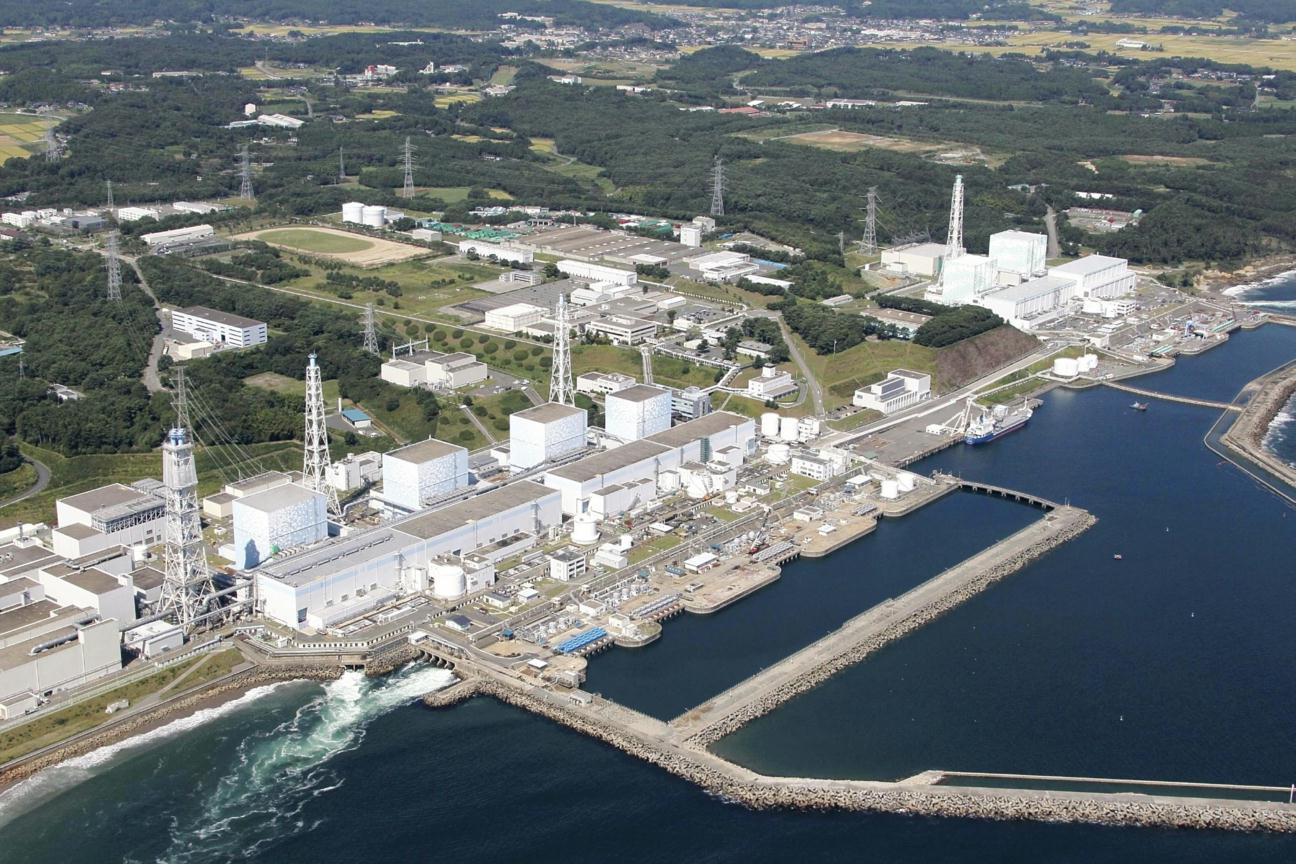
Fukushima Dai-ichi Nuclear Power Station before Mar. 11, 2011. Reactor units 1(right) - 4 (left) are seen in the foreground, units 5 and 6 are further in background (top left) (courtesy cryptome.org)
The fuel of three nuclear reactors melted down after the loss of coolant, a situation each should have incurred only once in 10,000 years or less, according to probabilistic risk assessment.

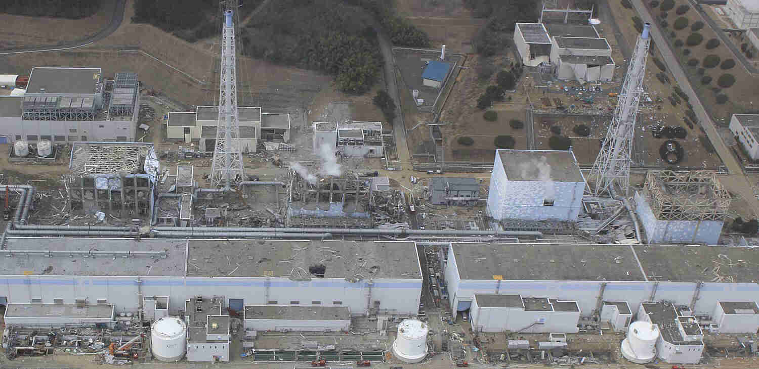
Fukushima Dai-ichi Nuclear Power Station after hydrogen explosions in the week after the Mar. 11, 2011, earthquake and tsunami. The explosions devastated the buildings of Units 1(right), 3 and 4. Unit 4 was not operating. The reactor incurred no fuel meltdown. By contrast, the fuel in units 1, 2 and 3 melted down and highly radioactive matter was released in amounts rivaled only by those released after the Chernobyl reactor accident (courtesy cryptome.org)
The meltdowns produced devastating hydrogen explosions that radioactively contaminated land, sea, and air to not yet fully comprehended extent. According to the report of the Japanese government to the International Atomic Energy Agency (page V-9) with the title "Report of Japanese Government to IAEA Ministerial Conference on Nuclear Safety - Accident at TEPCO's Fukushima Nuclear Power Stations (Jun.7, 2011)", about 78,200 residents in the immediate vicinity of the power station were evacuated and have only been permitted to return home for a few hours on occasion. A Japan Times article with the title "Tokyo faced evacuation scenario: Kan" published online on Sep. 19, 2011, reported from a recent interview with the Prime Minister of Japan at the time, Naoto Kan, that His Excellency contemplated the need to evacuate 20 million people from the greater Tokyo area at the height of the reactor crisis. He judged in the interview, “it was a crucial moment when I wasn't sure whether Japan could continue to function as a state.” No other statement could highlight the gravity of that emergency and the need of human organizations to perform adequately in extreme situations.

In the nine months that have passed since the accident, the operator of the Fukushima Dai-ichi Nuclear Power Station Tokyo Electric Power Company, has accomplished stable cooling of the still hot, molten mass of fuel and debris inside the highly compromised reactor buildings. The Japanese government is confronted with a massive environmental clean-up of the radioactive contamination. The challenges involved are unprecedented and daunting. The long-term effects on public health are unknown. Billions of yen will have to be spent to mitigate the damage.

Before this accident, nuclear power generation was considered safe in Japan. How was it possible that the most unthinkable scenario could happen three times in a row? How could the risk of nuclear power generation be that miscalculated?

Why we fail in our risk assessment has preoccupied generations of scholars. Charles Perrow's remarkable book with the title "Normal Accidents: Living with High-Risk Technologies" is still pertinent today. Perhaps, Professor Ewert's insights in the neuroethology of toads may contribute helpful insights.

Our brain takes decisions not unlike the toad's brain. In an area of cerebral cortex known as supplementary motor area for example, excitatory and inhibitory nerve cell inputs weigh in to promote or avert action (Jun and others, 2010; Lo and others, 2009).


Mandelbrot set in html5 (courtesy: Kostas Symeonidis, atopon.org).

Similar to a reverse Mandelbrot set, the fashion in which the nerve cells in our brain interact determines our behavior, which determines how we interact with each other, which determines the structure of human organizations.

Just as the brain consists of networks of nerve cells, human organizations consist of networks of people. Therefore, our mind cannot help, but act adhering to the brain's principles on the next higher level, that is inter-personal social interactions.

Each human enterprise comprises of members who wish to press forward with a promising idea, and members who demand moderation. In as much as the toad's brain, our brain balances cost against benefit. Factions within our organizations constantly struggle with each other over reward-versus-risk estimates. As long as checks and balances are built into our enterprises, we may succeed. By contrast, if one faction dominates the decision making process, either nothing can be achieved, or reckless risk-taking prevails. Through examination of small nerve cell assemblies in the brain, we may therefore attain an improved understanding on how human organizations must function to be successful.

The Fukushima reactor meltdowns represent an example that the unthinkable can happen. The presumption that the reactor design implemented at Fukushima was safe to withstand any fathomable seismic impact had dominated the views of the experts in the field since the inception of the commercial use of nuclear power. Japanese experts now readily acknowledge that the nonchalant attitude that has dominated the nuclear power industry in their country and its governmental regulatory body, the Nuclear and Industrial Safety Agency, (NISA), led to grave underestimations of risk for nuclear power reactors in earthquake and tsunami prone regions (NHKWorld News report with the title "Nuclear experts rethink their future" published online Sep. 20, 2011). Hierarchical report structures steeped in age-old traditions that permeate public and private endeavors like the Amakudari culture of Japan favor top-down instruction, vulnerable to perpetuate flaws in assumption.

In art, music and social relations, Japanese cherish harmony perhaps more than other cultures. Acclaimed medieval warriors like Miyamoto Musashi are equally known for their accomplished works of art, striving for harmony. Yet, like the nerve cells in our brain, our minds struggle every day with conflicting goals and ideas. Shinmen Mushashi represents an excellent example of this antagonism contained in the life of one person. This struggle must be allowed room for consensus to achieve the best possible outcome.

Nerve cell networks have evolved over eons, continuously improving our brain's design. Perhaps, the principles of neuroethology can help us improve the organization of human enterprises, and Professor Ewert's observations, models and simulations on the nerve cell networks underlying simple behaviors may be particularly informative, providing insights into the necessary components and strength of their relationships underlying successful social decision making. Hopefully, we can make use of such knowledge in the new year to develop a better understanding of risk that affects us all.

Acknowledgement
I thank Prof. J.-P. Ewert for teaching me about the neuroethology of toads. I thank Ranulfo Romo and Jeff Schall for sharing their insights into nerve cell mechanisms involved in decision making in the primate cerebral cortex. I am further indebted to simplyinfo.org and the commenters on www.scribblelive.com/Event/Japan_Earthquake5 for keeping me abreast on the latest developments in Japan.

Saturday, October 1, 2011

Whiskers, Manatees & The Mind


Harbor seal (Phoca vitulina),
courtesy L. Heafner. 
Unlike the follicles of common hair, the follicles of whiskers, also known as tactile vibrissae or sinus hairs, are complex sensory organs in which the hair is surrounded by a cushioning blood sinus and which are invested with thousands of innervated touch receptors of different type. Vincent (1913) described in great detail the intricate anatomy of the follicles of facial whisker in the rat, and she was the first to recognize the important role they play in tactile exploration and navigation.

Mammals with whiskers are not exclusively terrestrial. Pinnipeds possess whiskers on their snout.  Szymonowicz (1930) was the first to describe the follicular innervation of facial whisker follicles in the harbor seal Phoca vitulina. Recent behavioral studies provide astounding evidence that harbor seals can locate objects by their wake with their whiskers (Dehnhardt and others, 2001).

Not only carnivorous marine mammals seem to make good use of their whiskers that way. Notably, the herbivorous Florida manatee Trichechus manatus latirostris, a subspecies of the West Indian manatee, also appears to use whiskers for underwater exploration and navigation. Moreover, the long whiskers surrounding the mouth are employed for palpating and grasping plant matter when feeding. Florida manatees inhabit shallow, warm coastal waters. When the cold of winter arrives, the animals migrate south and up rivers into ponds and warm springs, where they congregate to forage and socialize.

Much we know about the Florida manatee brain, we owe to the ground-laying research of Professor Roger Reep from the University of Florida at Gainesville and his colleagues. Erica Goode described his work in her article with the title "Sleek? Well, No. Complex? Yes, Indeed." published online in The New York Times on Aug. 29, 2006. The article contains a slide show with Professor Reep's comments entitled "The Mind of The Manatee" which includes detailed photographs of the whiskers. Professor Reep co-authored an exhaustive book on manatees with the title "The Florida Manatee: Biology and Conservation".

The whiskers of the Florida manatee are densest on the face (2,000) with greatest density, roughly 600, in an area between the upper lip and the nose known as the oral disk (Reep and others, 2001).  Another 3,000 cover the remaining body (Reep and others, 2002). Sarko and others (2007) recently described the follicles of the manatee facial whiskers in anatomical detail. Although the facial whisker follicles receive at on average 50 myelinated nerve fibers per follicle almost twice the follicular innervation the whiskers of the rest of the body receive at 30, the heightened sense of touch comprising the entire body surface may represent a great advantage for navigating the dimly lit, muddy waters of the manatee's grazing grounds.

Considering their body size, manatees possess small brains (Reep and O'Shea, 1990). Unlike our heavily convoluted cerebral cortex, the manatee's is smooth showing only one fissure, homologous to our Rolandic fissure, running from top to bottom at the center of the cerebral hemisphere (shown in this NYT graphic with the title "A Sensitive creature"). Compared to ours, the manatee cortex seems thicker, and the gray matter differentiated in more layers. In addition, Professor Reep notes in Erica Goode's article condensations of nerve cells in the deep layers of somatic sensory cortex that may represent the whiskers topographically analogous to the barrels in mouse somatic sensory cortex (Woolsey and Van der Loos, 1970). Interestingly, Professor Reep also found such cell condensations in auditory cortex. The meaning of this finding is not yet understood.

Professor Reep further reports that manatees aptly localize sources of brief tone pips as low as 23 Hz in pitch. The cell condensations in auditory cortex may therefore suggest crossmodal processing of low frequency vibrations sensed with the whiskers. I discussed that rodent whiskers may detect low frequency ground motion in my post with the title "The Quest for the Infrasound Acoustic Fovea" published Oct 12, 2009. Without doubt, uncovering the nerve cell mechanisms underlying manatee behavior will provide exciting novel insights into the detection of low-frequency water motion by whiskered marine mammals and the processing of tactile input for underwater navigation.


If we peer through the above window preferably in the mornings EST, we may once in a while see manatees grazing and socializing in their habitat at Ellie Schiller Homosassa Springs Wildlife State Park, Florida, via their manatee cam (with permission, Susan Strawbridge, Ellie Schiller Homosassa Springs Wildlife State Park).

For close fullscreen examination of manatee whisker action, we must visit the park's manatee cam, which also provides a series of the ten most recent images for an improved sense of the action and detailed information on visitor activities at the park. An actual visit would be most exciting of course.

References
Related Posts

Saturday, May 1, 2010

The Beautiful Eimer's Organ

Maikäfer by H. Baluschek
May is the season of the Maikäfer in Germany, and moles become most active. Hence, in this month we celebrate Gustav Heinrich Theodor Eimer's arguably greatest discovery. Even Frau von Welt discussed it, though in fleeting. Theodor Eimer was born in Zürich, Switzerland, on Feb. 22, 1843, and passed away on May 29, 1898, in Tübingen, Southern Germany. His biography remains scant. After an extended search, I gleaned his birthday from the simple plaque on his grave pictured in this tour of Tübingen's Bergfriedhof. Plaque is a misnomer here. It consists of a rectangular steel plate with black lettering painted on gray rust-proofing. The simplicity does not come close to reflect his discovery.

Theodor Eimer aspired a career in comparative zoology and anatomy. He spent his junior faculty years as prosector at Julius-Maximillian's University, Würzburg, Germany, and moved on to hold a chair in zoology at Eberhard-Karl's University, Tübingen, where he led a productive life as an evolutionary biologist, struggling with Darwin's theory until his untimely death at the age of 55. His greatest scientific contribution, however, may have been as an early neuroethologist. Perhaps, it is this contribution that helped Tübingen's university and its three affiliated Max Planck Institutes develop into a hotbed of neuroscience today.

During his tenure in Würzburg, Theodor Eimer became the first to describe the discrete microscopic organ of touch that densely populates the tip of the nose of the European mole Talpia europaea. The organ is named in his honor. In his original publication (Eimer, 1871), he examined in great detail the structure of the nose, the distribution of the touch organs on the nasal skin, and the relationship of their density with the nose's use for palpation. Eimer sought to establish a connection between structure and function.

Eimer's organ fascinated me, ever since I saw one under the microscope in histological preparations Kenneth Catania showed me. Kenny is studying the North American star-nosed mole Condylura cristata. He counted roughly 25,000 Eimer's organs on these moles' nose appendages called rays. The organ consists of a minute skin papilla between a tenth and a fifth of a millimeter in diameter. At the papilla's core, a beautifully geometric constellation of nerve fibers with free endings is embedded with great symmetry in a conspicuous column of epithelial cells. Eimer (1871) saw two to three single nerve fibers, rising strait in the middle of the column and ending in the fifth layer under the stratum corneum that forms the hard top of the epidermis. The fibers extend short protrusions perpendicularly into each epithelial layer they traverse, where the protrusions end in 'buttons'. They are ringed by a circle of roughly 19 evenly-spaced nerve fibers, known as satellite fibers, whose protrusions point inwards. In addition, Eimer (1871) distinguished a separate set of nerve fibers with free nerve endings. By contrast to the fibers in the papilla's core, these travel obliquely toward the surface at the papilla's perimeter. Both sets are illustrated in his Fig. 2 of his 1871 publication, partially reproduced below:


His legend to this figure reads: Longitudinal section through the frontal surface of the mole's snout treated with the Gold-method. T touch cone, N nerve bundle, E epithelium, C corneum, magnification 400/1.

With improved histological techniques, a second touch receptor type, Merkel cell-neurite complexes, was found in the stratum germinativum at the bottom of the epidermis (darkest part in Eimer's figure), and a third, lamellated corpuscles of Vater and Pacini, was discovered in the stratum papillare of the dermis (white in Eimer's figure) underneath the Merkel cells (Halata, 1975).

Today, we still do not understand precisely how these receptors transduce touch into the electrical signals that the nerve fibers transmit to the brain. But we have learned much about the properties of touch, e.g. frequency and force, to which the receptors respond and how their responsiveness changes with prolonged stimulation. The receptors can be functionally distinguished based on these features. The nerve fibers with free nerve endings and the nerve fibers ending on Merkel cells adapt their responses to touch rapidly, whereas the nerve fibers ending in the lamellated corpuscles are considered slowly adapting.

Marasco and others (2006) were able to attribute different functions to Eimer's two sets of free-ending nerve fibers in the star-nosed mole and the coast mole Scapanus orarius. The authors furnish outstanding micrographs of the organ and its innervation, depicting Eimer's free-ending fibers as well as the Merkel cell-neurite complexes and the Vater-Pacini corpuscles. Using a histochemical marker for a protein known to be involved in the processing of pain, they were able to label the nerve fibers at the perimeter of the papilla, suggesting that they are nociceptive. That is, they respond to pain. By contrast, the fibers in papilla's core did not stain for the protein, suggesting that they are mechanoreceptive. These nerve fibers as well as the Merkel cell-neurite complexes are known to respond to local touches with great sensitivity, whereas the Vater-Pacini corpuscles are highly tuned to the frequencies of dispersed vibrations. Eimer's organ, therefore, forms a receptor complex, integrating pain receptors as well as three fundamentally different types of touch receptors which preferentially respond to either skin indentations or vibrations. The follicles of whiskers, also known as vibrissae or sinus hairs, and the push rods in monotremes (Proske and others, 1998) represent the only other known discrete structures in the skin that combine three mechanoreceptor types.

The Eimer's organs on the nose may be the mole's main tool with which the animal can capture a refined picture of its underworld.  Catania and Kaas (1995) have shown that the nose of the star-nosed mole is mapped in multiple topographic representations on an extraordinarily large swath of cerebral cortex that processes touch. Discrete morphological modules of nerve cells that are clearly discernible in histologically stained sections represent each ray in the same order as they surround the nose. This topographic morphological representation of the sensory periphery is similar to that of the facial whiskers by cytoarchitectonic modules called barrels in the rodent cerebral cortex. I touched on whiskers and barrels in my posts dated Oct. 12, 2009, and Dec. 24,  2008.

To date, two complete cortical maps of the nose with its rays have been found in the brain of the star-nosed mole. There may be more. The nose's disproportionate representation in cerebral cortex is suggestive of a fovea for nose touch in the mole's somatic sensory system (Catania, 1995).


Already Professor Eimer recognized the importance of the mole's nose to the behavior of the species. He begins the introduction to his 1871 publication with the statement: "The mole's snout must be the seat of an extraordinarily well developed sense of touch, because it replaces almost entirely the animal's sense of face, constituting its only guide on its paths underground." He estimated that the nose of the European mole was covered with more than 5,000 Eimer's organs which were invested with 105,000 nerve fibers. He took the abundance of sensory innervation to affirm his contention that the nose's touch must represent the moles dominant facial sense. Professor Eimer ends his report with the assertion that his interpretation is entirely consistent with the common knowledge of his time. His last sentence reads: “This enormous richness of innervation easily explains the well-known fact that already a light blow to the snout kills the mole instantly.”

Roughly 130 years after Professor Eimer's discovery, Catania and colleagues accomplished to record striking behavioral evidence in favor of his conclusion, using a high-speed camera that the eminent professor could hardly have imagined  (Catania and Remple, 2004).  Professor Eimer would have been fascinated, watching this footage. Moles with the help of their Eimer's organs may be perfectly poised to detect seismic vibrations.

References
Related Posts


-->

Addendum
Impressively shown by the two Reuters news clips below, high-speed video cameras have developed into invaluable tools in the study of animal behavior. Their application range from the examination of bat flight
to feline drinking, providing new insights propelling engineering design (11/12/10).

Thursday, January 28, 2010

Cell Conversion: Potential Solution for Neurodegenerative Diseases

About 10 percent of the genes in the human genome encode transcription factors. Transcription factors are proteins that bind to DNA, controlling gene expression. Notably, they control the precisely-timed differentiation of stem cells into specialized tissues during the development of organisms.

In an article published online in the journal Nature this week, Marius Wernig and colleagues at Stanford University demonstrate that the expression of only three transcription factors, that is Ascl1, Pou3f2 and Myt1l, is needed to convert cultured fibroblasts, that is undifferentiated connective tissue cells, into nerve cells (Vierbuchen and others, 2010). The cells were shown to produce action potentials, i.e. the electrical discharges that propagate along their output processes called axons, and formed functional connections, known as synapses, with other nerve cells.

Although the conversion has been successfully carried out only in a dish with developing mouse tissue and only the most essential nerve cell functions have been demonstrated, the method does not utilize stem cells and still may bear great potential for therapies, seeking to replace lost nerve cells in neurodegenerative diseases, e.g. Parkinson's disease.

Addendum

References
Related Posts


Thursday, December 3, 2009

H.M.'s Brain

The philosopher Martin Heidegger reasoned in his treatise "Sein und Zeit" that our sense of time confers our sense of being (Heidegger, 1976). Henry Gustav Molaison could attest to that. In order to relieve Henry from severe bouts of epileptic seizures, a part of his temporal lobes had been removed when he was in his late twenties.

The seizures subsided. However, as an unwanted consequence of the surgery, he had lost essential elements of his memory. He could not remember who he was and had to discover his identity anew every day. One day late in his adult life, Henry was caught curiously examining his face in a mirror. He called out in amazement, "I am not a boy!" In Heidegger's words, Henry Molaison had lost his sense of time and, with that, his sense of being.

As patient H.M., Henry volunteered for scientific research throughout his life, becoming one of the most intensely examined cases in psychology. He willed his brain to research. Yesterday a year ago, he passed away of respiratory complications at the age of 82. On the first anniversary of his passing, the histological processing of his brain was begun at the University of California San Diego. The university's Brain Observatory provides live online footage of the sectioning of the frozen whole brain on a cryotome until Dec. 4, 2009. The brain is cut coronally, that is in the transverse plane. Seventy micrometer-thin sections are collected for histology at equally-spaced intervals. At the speed maintained thus far, we shall reach the site of surgery tomorrow.

The ensuing histological examination of his brain will complete Henry's scientific journey as he willed. The findings may provide important insights into the histochemistry of memory, constituting the keystone in the arch of knowledge that H.M. allowed us to build.

Related Posts

Addenda
  • At 5:00 am (CST), we are in the midst of the temporal lobe of H.M.'s brain. If you look closely at the bottom of the brain (ventral, on the left hand side of the observer), you see cauliflower-shaped protrusions on both sides. These are the temporal lobes. There is a void inside the structures. Parts of the hippocampus seem absent (12/04/09).
  • At about noon, in section 1672 (46,344 micrometers), we can see a cavity in left lingual gyrus (in the hemisphere closest to the observer) (12/04/09).
  • At 5:00 pm (CST), we are entering the occipital lobes; at 10:00 pm we passed the middle of the occipital lobes (12/04/09). 
  • We should be able to detect signs of nerve cell and nerve fiber degeneration in the brain regions that were connected to the brain tissue that was removed or damaged otherwise during surgery, particularly in the structures of the limbic system. Degenerating nerve fibers can be rendered decades after the damage and ameboid microglia may reside in the affected locations (Miklossy and others, 1991). According to von Monakow's concept of diaschisis, the affected regions can be far-flung and distant from the site of immediate damage, comprising the opposite cerebral and cerebellar hemispheres. That is why H.M.'s whole brain needed to be sectioned, and 2,401 sections were collected (12/05/09).
  • The footage covering the sectioning does not seem to be available on The Brain Observatory's site at this time. You may find a CBS report broadcast Dec. 3, 2009, instructive (01/01/10):
  • Brain sections are prepared for histology with two distinctly different methods. One way requires that the tissue is embedded in hot paraffin wax or plastic. The tissue is sectioned at room temperature on a bench top with a microtome or ultramicrotome, respectively. Paraffin embedding is widely used in routine pathology. Plastic is used to embed specimen to be cut into ultra-thin sections for electron microscopy. The other method of tissue preparation requires that the specimen are frozen. Frozen tissue has to be stored and sectioned at temperatures below 0 ℃.

    With either method of preparation, the tissue is commonly first soaked in solutions of formaldehyde, glutaraldehyde or a mixture of both. The aldehydes help bond cellular protein, preserving cellular structure during the further processing steps. H.M.'s brain was then soaked in highly concentrated sucrose solutions and embedded in dextran. Sucrose and dextran are sugars. The sucrose prevents ice crystals from forming in the cells when the tissue is frozen. The growing crystals would otherwise burst the cells. The dextran helps preserve the integrity of the sections, preventing tissue from breaking off the perimeter of the specimen during cutting. Once the brain is fixed and cryo-protected, it can be cut into sections that are only tens of micrometers thick on a microtome on which only the stage holding the brain is kept at subzero temperatures. The sections are picked up from the microtome's knife with a brush and can be processed freely floating in solutions containing the substances needed for the chosen histological staining procedure.

    There are numerous histological staining procedures. Most comprise chemical reactions. Others involve antibodies that bind specifically to cellular proteins or fluorescent dyes that fill particular cellular components. Among the oldest procedures is the silver impregnation method named after  Camillo Golgi, rendering the cell bodies of nerve cells and a good part of their processes (dendrites) and protrusions (dendritic spines). This method helped Ramón Cajal to divide the cerebral cortex into six layers, a distinction that is still used today. Golgi and Cajal were awarded the Nobel Prize for their contributions. The most commonly employed methods, however, are the Nissl stain for cell bodies and the myelin stain that stains the fatty sheeth around nerve cell fibers in the brain's white matter. These methods will certainly be used with some sections of H.M.'s brain. After staining, the sections are mounted on glass slides, dehydrated in solutions of increasing alcohol concentration, embedded in a clear organic solvent-based glue and covered with thin glass coverslips.

    The methods of tissue preparation described above cannot be used when the procedures require that fixation-sensitive cellular proteins remain functional or when water-soluble, diffusible substances are to be localized in the tissue. In that case, the tissue must remain unfixed and immersion in sucrose cannot be used. The tissue has to be shock frozen at -70 ℃ and lower, and the sections must be kept frozen during sectioning. To fulfill this need the whole microtome is encased in a refrigerated cabinet. The instrument is then called a cryostat. Ten or 20 micrometer-thick sections are cut and picked up on chilled glass slides and quickly dried on a hotplate outside the cabinet. The subsequent procedures must consequently be carried out on the slide-mounted sections. This method is used for the autoradiography of diffusible radioactively-labeled tracers. For autoradiography, the sections are exposed to x-ray films or radiation-sensitive semiconductor chips that produce gray-scale images of the distribution of the tracer in the sections. The optical densities in the images called autoradiograms are proportional to the concentration of the tracer in the tissue and can be measured.

    In order to trace metabolites in whole bodies, Sven Ullberg and colleagues at Uppsala University, Sweden, engineered a microtome sufficiently sturdy to cut thin sections through the whole body of small animals. The microtome was placed it in a chest freezer with windows in the lid (Larsson and Ullberg, 1981). The instrument is called a whole-body cryotome and was commercially available from the Swedish company LKB. I was privileged to use a LKB cryotome 30 years ago for functional imaging studies (Melzer, 1984). The picture below shows a whole rat embedded length-wise in dextran and frozen in liquid nitrogen at -173 ℃. The dextran block with the rat is mounted on the sliding stage of the cryotome for sectioning.
    The block of dextran with the rat resembles that with H.M.'s brain in the footage of the Brain Observatory which misled me initially. But as I pointed out, H.M.'s brain was fixed and protected with sucrose. Only the microtome stage had to be cooled. No freezer chest was needed. Jacopo's hands stayed warm.

    One great advantage of whole body sectioning is that the sections render the organs in their actual position inside the body. The picture below shows a the block surface of the head of a sandrat embedded in dextran and cut transversely at the level of the midbrain.
    The structure at the center is the brain. The glassy cavities on both sides of the brain are the inner and middle ears. The middle ear cavities of sandrats are remarkably enlarged compared with other rodents, serving as a low pass filter. The animal had been exposed to sound for functional imaging. Brain activation images are shown in my post entitled "The Quest for the Infrasound Acoustic Fovea" and dated Oct. 12, 2009.

    Since the brain was preserved accurately as it was situated in the scull, we were able to pinpoint brain structures with stereotaxic precision, a goal that may be achieved with structural magnetic resonance imaging today. That method did not exist then.

    In addition to functional imaging, fresh tissue tissue sections permit us to examine the distribution of radioactively-labeled ligands that bind to specific receptors on cell surface or probes that identify sequences of genetic code. These procedures can not be applied to the sections of H.M.'s brain. However, even with the histological techniques available a treasure of knowledge will be uncovered from the brain that Harvey Molaison kindly willed.

    In the past three decades most departments of anatomy have been transformed into departments of cell biology for good reason. However, gazing through a microscope at the multitude of diversely shaped nerve cells in a Golgi-stained section through cerebral cortex still remains as fascinating as gazing through a telescope at the myriad of stars in the night sky. The neuroanatomical methods that make this experience possible resemble in many ways forms of art, though rather of an artisan than of an artist. Perhaps, the work on Harvey's brain will rekindle interest in this type of expertise, paving the way for a renaissance of neuroanatomy (01/02/10).
  • The Allen Institute for Brain Science in Seattle, WA, compiled the first comprehensive atlas of local gene expression in the mouse brain five years ago and has now released a similar atlas for the human brain (04/13/2011):

References


Wednesday, November 4, 2009

Constantin von Monakow & Brain Plasticity

Constantin von Monakow was born in Bobresovo, Poland at that time, on this day in 1853.  He was to become one of the most eminent neuroanatomists, neurologists and psychiatrists of his age.  Below I attempt to highlight some of his achievements, relying mainly on Mario Wiesendanger's informative article in the Comptes Rendus Biologies (Wiesendanger, 2006).

Von Monakow spent most of his life in and near Zürich, Switzerland, where his family moved in his youth. He studied medicine, worked with Eduard Hitzig at one of the first psychiatric research clinics in continental Europe, the Burghölzli. The principal investigators at the clinic led by Auguste Forel had fully recognized the role of cerebral organization in mental illness.  Hitzig and Gustav Theodor Fritsch were among the first to localize function in the cerebral cortex with electrical stimulation electrodes.  Hitizig hired von Monakow as assistant and taught him histological methods.  At a brief visit with the famous anatomist Bernhard von Gudden in Munich, he familiarized himself further with silver impregnation methods that permitted him to visualize degenerating nerve fibers. Injury to brain tissue, precipitated by hemorrhagic bleeding, stroke or trauma, results in the degeneration of the nerve cell connections between the affected location with other parts of the brain.  Tracing degenerating nerve fibers, therefore, could be used to examine pathways among brain structures.

Von Monakows independent research would begin under small and unusual circumstances as attending physician at St. Pirminsberg, a psychiatric asylum near Bad Ragaz, a spa not far from Zürich.  One day, while inspecting the premises, he discovered a brand new microtome stowed away in an unused closet.  He knew how to use the microtome and turned the pantry into a small histological laboratory to get a series of experiments off the ground that would uncover the brain's visual pathway.  The faculty of the University of Zürich accepted the publication of the results of these experiments as qualification for the right to teach (habilitation), and he started to give lessons at the university without salary, while attempting to maintain a private practice for neurology and general medicine in the city.  The practice barely supported him. Yet he managed to continue his research in a small laboratory he sat up for himself.

Eventually in 1894, von Monakow received a call from the University of Innsbruck, Austria, to assume a chair in psychiatry.  The offer compelled the government of Zürich to make a counterproposal which von Monakow happily accepted, although the appointment was only at the level of associate professor. Despite the lower rank, the new post permitted him to continue his research in his laboratory, which became known as the Brain Institute and serve as the director of a psychiatric policlinic.  Finally, 17 years after he graduated from medical school, he had garnered a stable position, securing financial support for his research and a stable income.

Von Monakow worked full-time at the university until his retirement at the age of 70, and continued another four years as honorary professor and director of his laboratory, which became known as the Brain Institute. During his years as professor, he maintained an active journal club, enjoying regular visits from a number of eminent scientists and physicians. Among others, the fourth director of the Burghölzli, Auguste Forel, and the famed neuroanatomist and neurologist Constantin von Economo were in attendance as much as the founder of psychotherapy, Carl Gustav Jung, whose long-hidden self-analysis, known as The Red Book, has just been published. 

Constantin von Monakow passed away in 1930 at the age of 77, and his former responsibilities were divided among several successors.  In 1962, Konrad Akert succeeded in recreating Von Monakow's Brain Institute as The Institute for Brain Research, which has remained a vibrant site for world-class neuroscience research to the day. 

Constantin von Monakow was a prolific writer and published a long list of scientific articles, book chapters, books, hand books and anatomical brain atlases.  His work entitled "Die Lokalisation im Grosshirn und der Abbau der Funktion durch kortikale Herde" (Localization in the Cerebral Cortex and Loss of Function Produced by Cortical Lesions) arguably became one of his most notable contributions.  In this book, he laid out comprehensively the principles of chronogenic localization and diaschisis.

Von Monakow's observations suggested that the cortical and subcortical components of a brain pathway cooperate in precisely timed sequence to synergistically effect coordinated brain function, though the components may not necessarily be situated near each other.  He called the cooperative, yet distributed localization of brain function chronogenic localization.  His concept ran counter to the widely-held belief at the time that brain function was localized in circumscribed centers dedicated to particular tasks.  Phrenology represented the most extreme and popular variation of that idea.  History would prove von Monakow right.  The functional brain imaging studies conducted today commonly render multiple loci of activation in cerebral cortex when the participants execute a task, confirming that brain functions are indeed carried out by distributed nerve cell networks involving a number of cortical areas.

For example, using functional magnetic resonance imaging, my colleagues and I observed numerous foci of activation scattered across cerebral cortex when people with severe visual disability read Braille with their fingertips (Melzer and others, 2001). Intriguingly, cortical areas in the occipital lobes were involved that process visual information in sighted people. The animation below shows these foci in a 36 mm slice through cerebral cortex. The poles of the occipital lobes point to the bottom at the finish.


As another significant finding, von Monakow observed that acute damage to one component of a brain system immediately depressed the function of the system's unharmed components, disabling their coordinated cooperation.  He called this immediate loss of adequate control over integrated brain function diaschisis.

Von Monakow noted furthermore that with time functionality would return, though a residual deficit almost always remained. Differences in the timing of the recovery of various aspects of the lost function would bare its chronogenic localization.  The recovery would help identify each component's contribution to the integrated function.  Nerve cell plasticity would permit the unharmed components to reorganize and reintegrate. However, he could only speculate on the underlying cellular mechanisms.

Von Monakow proposed that the increased use of existing, undamaged collateral nerve cell connections within cerebral cortex stimulated the development of new connections, recruiting nerve cells in other cortical areas for the recovery of lost function. Furthermore, he recognized that exercise benefited recovery, suggesting that exercise-related stimulation facilitated the growth of new, more extensive nerve cell connections. As a consequence novel behaviors would be learned to compensate for residual deficits.

In own research, my colleagues and I observed that nerve cells in the vicinity of a stroke lesion in the cerebral cortex respond to sensory input only at short latency in the days after the infarct.  By contrast, responses at long latency were suppressed (Melzer and others, 2006). Inputs from intracortical nerve cell connections are thought to drive the long latency responses.  The observed suppression is entirely consistent with von Monakow's diaschisis.

Von Monakow's concept of distributed nerve cell networks underlying brain function was astoundingly modern.  His ideas of recovery of function were profoundly forward looking.  More than a century after their inception, his ideas are still inspiring intense research.

References
  • Melzer P, Maguire MJ, Ebner FF (2006) Rat barrel cortex as a model for stroke analysis. Soc Neurosci Abst:583.13.
  • Melzer P, Morgan VL, Pickens DR, Price RR, Wall RS, Ebner FF (2001) Cortical activation during Braille reading is influenced by early visual experience in subjects with severe visual disability: a correlational fMRI study. Hum Brain Mapp 14:186-195.
  • Monakow von C (1914) Die Lokalisation im Grosshirn und der Abbau der Funktion durch kortikale Herde. J.F. Bergmann, Wiesbaden.
  • Wiesendanger M (2006) Constantin von Monakow (1853-1930): a pioneer in interdisciplinary brain research and a humanist. C R Biol 329:406-418.
Related Posts


Thursday, October 22, 2009

Absolutes, Relatives, Brain Imaging & Steroids

The functional brain imaging methods most commonly used in humans today are functional magnetic resonance imaging (fMRI) using blood oxygen-level dependent (BOLD) signals and the subtractive water method with positron emission tomography (PET). Both procedures record changes in local cerebral blood flow from a baseline. Local cerebral blood flow is associated with energy demands of activated nerve cells. The cells consume glucose sugar and oxygen to process information which cannot be stored in the brain and must be supplied on demand with the blood stream. Hence, blood flow increases with increased nerve cell activity. In the simplest conception of both procedures known as block design, measurements acquired over several minutes of mental activation are compared with measurements acquired during equivalent epochs of rest. The statistically significant difference is considered the result of cerebral activation.

In conventional fMRI, blood oxygen-level dependent signals are used to detected changes in blood flow (Ogawa and others, 1993). With the subtractive water method (Fox and others, 1984), water labeled with 15O, a radioactive positron-emitting isotope of oxygen, is used as tracer that freely diffuses into the brain tissue. The local concentration of the tracer is commensurate with local blood flow and can be imaged in a PET scanner. However, calibration for absolute blood flow is wrought with difficulty and has not found wide-spread application. Because the blood flow is not quantified, the differences between the compared mental states remain relative. That is, cerebral activation is usually expressed in percent difference from the state used as reference or in units of statistically significant difference (statistical parametric mapping).

By contrast, the cerebral glucose consumption is more directly related to nerve cell activity than cerebral blood flow. The deoxyglucose method of Sokoloff and others (1977) permits us to measure the local cerebral rate of glucose utilization. Deoxyglucose is an analogue to glucose that accumulates in the brain tissue commensurate with glucose consumption. Tagging deoxyglucose with 18F, a radioactive positron-emitting isotope of fluorine, the tracer's accumulation in the brain can be imaged with PET. The [18F]fluorodeoxyglucose method, therefore, provides a snapshot of the brain's energy consumption (Reivich and others, 1979). Although this snapshot needs 90 minutes to develop because of the tracer kinetics involved, the procedure constitutes an indispensable tool for the detection of long-term, pseudo-stationary changes in absolute cerebral metabolic activity as a consequence of disease or trauma. Below, I discuss one example.

After the collapse of the regime of Nicolae Ceauşescu at the end of 1989, U.S. parents began to adopt children from Romanian orphanages. The children had been kept in circumstances of great depravity, producing profound behavioral problems similar to autism (American RadioWorks report, 2006). Visiting scientists reported behavioral patterns resembling those the eminent American psychologist Harry Harlow had so aptly described in primates raised in isolation and with surrogates.


Although the adoptees were brought to the U.S. at very young age, some developed cognitive and behavioral differences, including impulsive reactions as well as attention and social deficits, in the years after their arrival.

Research at the orphanages provided evidence that the children had persistently augmented levels of cortisol n their blood stream as a result of the severe stress they endured (Carlson and Earls, 1997). Cortisol is a known steroid stress hormone produced in the adrenal glands and can fundamentally affect brain maturation. The hormone suppresses the activity of glia. A type of glia, astrocytes, helps regulate the extracellular glutamate concentration. Glutamate constitutes the most prevalent excitatory neurotransmitter in the brain, playing a major role in the stabilization of connections between nerve cells during brain maturation. Elevated concentrations of extracellular glutamate can trigger pre-programmed cell death known as apoptosis, otherwise occurring only during early stages of brain development. Presumably, the orphans' excessive stress-related exposure to cortisol led to modifications of nerve cell networks, underlying the children's behavioral differences. Imaging the brain's energy consumption provided a method to uncover whether and where nerve cell activity changed in cerebral cortex as a consequence of the children's stay in the orphanages. 

Using the fluorodeoxyglucose method, Chugani and others (2001) could show that the use of glucose was drastically reduced in the cerebral cortex of the orphans enrolled in the study, particularly in temporal and prefrontal cortical areas and in structures of the limbic system, notably the amygdala. The cortical regions are involved in executive functions and short-term memory crucial for social behavior and affect. The amygdala play an important role in fearful reactions. The observed reductions in energy consumption could not have been detected with the standard fMRI or PET procedures discussed above. The fluorodeoxyglucose method, hence, constitutes the procedure of choice when the fundamental metabolic state of the brain is in question.

Addendum
  • Take some time and listen to this show on National Public Radio's This American Life with the title "Unconditional Love". The first half of the show is about an orphaned Romanian boy adopted by an American couple at the age of eight. It demonstrates in great clarity the at times overwhelming difficulties the family faced to remedy important steps of personality development that were missed early in the boy's life. Finally, the challenges were overcome with passion and a professional attitude. It is reassuring to find out that success is possible (10/23/10).

References

Wednesday, September 30, 2009

Echolocation, Science & Power

The Italian scientist Lazzaro Spallanzani  attained fame beyond his country already in his life time. He studied philosophy at the University of Bologna, became member of the clergy, and taught logic, metaphysics, and Greek at the universities of Reggio, Modena and and eventually assumed the chair in natural history at Pavia. His studies contributed profoundly to the understanding of a broad range of natural phenomena spanning geology, biology and physiology. Late in his career, he became fascinated with bats, wondering how they could navigate so elegantly in full darkness complicated environments wrought with obstacles.

Spallanzani proved himself as an observant experimenter. He attacked his question methodically with a series of tedious behavioral experiments, systemically ruling out one sense after another. He observed bats flying skillfully passed nooks and crannies in a L-shaped basement. Occlusion of the eyes did not appear to degrade the bats' performance, neither did covering the skin with a paste nor occluding the nose. His findings were first published as a collection of letters by Anton-Maria Vasalli (1794). A few years later, the Swiss zoologist Charles Jurine observed that occlusion of the ears rendered bats entirely disoriented. Spallanzani confirmed this observation, but was unable to explain how bats would use hearing for navigation. He had speculated earlier that the animals perhaps possessed a sixth sense unbeknownst to human kind.

Professor Spallanzani's hypothesis was met by strong and powerful resistance in the scientific community. One of the most eminent zoologists of his time, Georges Cuvier, argued against the validity of the experiments (Cuvier, 1795).  Interestingly, Cuvier's arguments were entirely based on conjecture. He did not conduct a single experiment to disprove Spallanzani's results. Instead, he appealed to common knowledge. He reasoned that everybody knows that bats, in as much as blind people, orient themselves with the sense of touch.

History would prove Cuvier wrong on both accounts. Neither bats (Griffin, 2001) nor blind people ( Wall Emerson and Ashmead, 2008) use their sense of touch for navigation in space. However, Cuvier's reputation was domineering. His influence on science was overarching. Roughly for the next century and a half researchers devoted their attention on the bats' sense of touch, until the Americans Donald R. Griffin and Robert Galambos and the Dutchman Sven Dijkgraaf discovered echolocation. That is, they unequivocally demonstrated that bats use the echos of ultrasound they emit to navigate their environment in flight and catch prey (Griffin, 2001). Griffin wrote an informative popular book about his findings entitled "Listening in the Dark: The Acoustic Orientation of Bats and Men". Vigorous research continues to the day to elucidate the nerve cell mechanisms that underlie this fascinating behavior.


I was exposed to some aspects of bat research when I was a student. The processing of ultrasound frequencies used for echolocation constitutes a prominent feature in the auditory pathway of echolocating bats (Neuweiler and others, 1980). My pilot study helped visualize this prominence with a functional imaging method (Melzer P, 1985).

The colors in the picture show ultrasound-related activation in a transverse slice through the brain of an echolocating bat. The ear on the opposite side was exposed to sound pips of this bat's individual echolocation frequency. Two regions in the auditory midbrain known as inferior colliculus (white circle) responded to the ultrasound most prominently.

Spallanzani's observations were influenced by the fact that he used pipistrelle bats (Pipistrellus pipistrellus) for his study. They emit their echolocation calls through the mouth, and his attempts to occlude the mouth noticeably compromised navigation. Had he used the more common horseshoe bat (Rhinolophus ferrumequinum), he would have been surprised to discover that the nose was doing the job.

Addendum
  • The co-discoverer of bat echolocation Robert Galambos, PhD, MD, passed away a month ago at the age of 96.  Douglas Martin provides a concise summary of his career in The New York Times today with the title "Robert Galambos, Neuroscientist Who Showed How Bats Navigate, Dies at 96".  He was a profound experimentalist. In an inseminating recent paper (Galambos, 2003), he described four elegant experiments that demonstrate the principles of empirical neuroscience in most illustrative fashion (07/16/10).
References
  • Cuvier G (1795) Conjectures sur le sixiéme sens qu'on a cru remarquer dans les chauve-souris. Mag. Encyclopéd 6:297-301.
  • Galambos R (2003) Four favorite experiments and why I like them. Int J Psychophysiol 48:133-140.
  • Griffin DR (2001) Return to the magic well: Echolocation behavior of bats and responses of insect prey. BioScience 51:555–556.
  • Griffin DR (1958) Listening in the dark: The acoustic orientation of Bats and men. Yale Univ Press.
  • Melzer P (1985) A deoxyglucose study on auditory responses in the bat Rhinolophus rouxi. Brain Res Bull 15:677-681.
  • Neuweiler G, Bruns V, Schuller G (1980) Ears adapted for the detection of motion, or how echolocating bats have exploited the capacities of the mammalian auditory system. J Acoust Soc Am 68:741-753.
  • Vasalli A-M (1794) Lettere sopra il Sospetto di un Nuovo Senso nei Pipistrelli . . . Con le Risposte dell’Abate. Stamperia Reale (Torino).
  • Wall Emerson R, Ashmead D (2008) Visual Experience and the concept of compensatory spatial hearing abilities. In: Blindness and brain plasticity in navigation and object perception (Rieser JJ, Ashmead DH, Ebner FF, Corn AL, eds). Taylor & Francis (New York):pp367-380.
Meet Bert the kitchen bat!